Convenience & Accuracy
Highest quality imaging services, highly knowledgeable technologists, fast & accurate without compromising the quality of our services. Exams are done in a patient friendly environment, with convenient scheduling option
Reliability & Experience
Certified radiologists by The American Board of Radiology, Licensed through Medical Board of California and certified through State of California Department of Public Health with over 30 years of experience.
Our highly experienced technologists are certified and credentialed.
Efficient Interaction & Accessibility
Multilingual staff keeping the consultation between Radiologists and Physicians efficient. Digital technology providing RAD reports & images to physicians through secured site as they become available.
Welcome!
JUST X-RAYS welcomes all insured, non insured patients, Medicare, and Crossover Medical patients, Military recruits through USMEPCOM, and Veterans with a valid form of referral mandated by American Medical Association, California Medical Association and Centers for Medicare & Medicaid Services
Why Us?
 More than 10 years of experience
Radiology facility with a heart for medical care of the community, providing accurate diagnosis at the most affordable price with the most convenient scheduling
Info Center
- X-ray of Bone
- X-ray of Chest
- Ultrasound General
- Ultrasound Abdomen
- Ultrasound Breast
- Ultrasound Carotid
- Ultrasound Scrotum
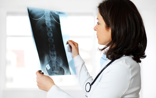
X-ray of Bone | Close it
A bone x-ray makes images of any bone in the body, including the hand, wrist, arm, elbow, shoulder, foot, ankle, leg (shin), knee, thigh, hip, pelvis or spine. Read more
A bone x-ray makes images of any bone in the body, including the hand, wrist, arm, elbow, shoulder, foot, ankle, leg (shin), knee, thigh, hip, pelvis or spine. Read more
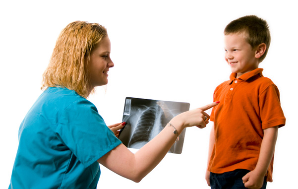
X-ray of Chest | Close it
The chest x-ray is the most commonly performed diagnostic x-ray examination. A chest x-ray makes images of the heart, lungs, airways, blood vessels and the bones of the spine and chest. Read more
The chest x-ray is the most commonly performed diagnostic x-ray examination. A chest x-ray makes images of the heart, lungs, airways, blood vessels and the bones of the spine and chest. Read more
Sonohysterography | Close it
Sonohysterography, also known as saline infusion sonography, is a special, minimally invasive ultrasound technique. It provides pictures of the inside of a woman's uterus. A Doppler ultrasound study may be part of a sonohysterography examination. Doppler ultrasound is a special ultrasound technique that evaluates blood as it flows through a blood vessel. Read more
Sonohysterography, also known as saline infusion sonography, is a special, minimally invasive ultrasound technique. It provides pictures of the inside of a woman's uterus. A Doppler ultrasound study may be part of a sonohysterography examination. Doppler ultrasound is a special ultrasound technique that evaluates blood as it flows through a blood vessel. Read more
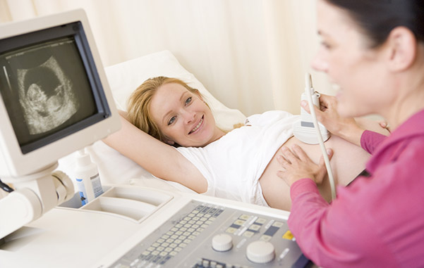
Ultrasound General | Close it
Ultrasound imaging, also called ultrasound scanning or sonography, involves the use of a small transducer (probe) and ultrasound gel to expose the body to high-frequency sound waves. Ultrasound is safe and painless, and produces pictures of the inside of the body using sound waves. Ultrasound examinations do not use ionizing radiation (as used in x-rays). Because ultrasound images are captured in real-time, they can show the structure and movement of the body's internal organs, as well as blood flowing through blood vessels. Ultrasound imaging is a noninvasive medical test that helps physicians diagnose and treat medical conditions. Read more
Ultrasound imaging, also called ultrasound scanning or sonography, involves the use of a small transducer (probe) and ultrasound gel to expose the body to high-frequency sound waves. Ultrasound is safe and painless, and produces pictures of the inside of the body using sound waves. Ultrasound examinations do not use ionizing radiation (as used in x-rays). Because ultrasound images are captured in real-time, they can show the structure and movement of the body's internal organs, as well as blood flowing through blood vessels. Ultrasound imaging is a noninvasive medical test that helps physicians diagnose and treat medical conditions. Read more
Ultrasound Abdomen | Close it
An abdominal ultrasound produces a picture of the organs and other structures in the upper abdomen.
A Doppler ultrasound study may be part of an abdominal ultrasound examination.
Doppler ultrasound is a special ultrasound technique that evaluates blood flow through a blood vessel, including the body's major arteries and veins in the abdomen, arms, legs and neck. Read more
Ultrasound Breast | Close it
Ultrasound imaging of the breast produces a picture of the internal structures of the breast.
Doppler ultrasound is a special ultrasound technique that evaluates blood flow through a blood vessel, including the body's major arteries and veins in the abdomen, arms, legs and neck.
During a breast ultrasound examination the sonographer or physician performing the test may use Doppler techniques to evaluate blood flow or lack of flow in any breast mass. In some cases this may provide additional information as to the cause of the mass. Read more
Ultrasound Carotid | Close it
An ultrasound of the body's two carotid arteries, which are located on each side of the neck and carry blood from the heart to the brain, provides detailed pictures of these blood vessels and information about the blood flowing through them.
A Doppler ultrasound study is usually an integral part of a carotid ultrasound examination.
Doppler ultrasound is a special ultrasound technique that evaluates blood flow through a blood vessel, including the body's major arteries and veins in the abdomen, arms, legs and neck. Read more
Ultrasound Scrotum | Close it
Ultrasound imaging of the scrotum provides pictures of the testicles and the surrounding tissues of a man or a boy.
Ultrasound imaging, also called ultrasound scanning or sonography, involves the use of a small transducer (probe) and ultrasound gel to expose the body to high-frequency sound waves. Ultrasound is safe and painless, and produces pictures of the inside of the body using sound waves. Ultrasound examinations do not use ionizing radiation (as used in x-rays). Because ultrasound images are captured in real-time, they can show the structure and movement of the body's internal organs, as well as blood flowing through blood vessels.
Ultrasound imaging is a noninvasive medical test that helps physicians diagnose and treat medical conditions. Read more
Using innovative, high quality digital imaging solutions to bring an outstanding experience to you