Our Services
We provide a wide range of X-Ray and Ultrasound services for adults and infants, in following categories:
X-RAY:
- > Head & Neck
- > Chest & Abdomen
- > Spine
- > Upper Extremities
- > Lower Extremities
ULTRASOUND:
- > Small Parts
- > Abdomen
- > Pelvic
- > Vascular
- > Soft Tissue
Physicians can download a requisition form for a detailed list of services provided from the “Links & Forms” tab.
Please note, in compliance with the Centers for Medicare & Medicaid Services, you may only receive these services if you are referred to us by a Medical Doctor, or authorized Medical practitioner.
JUST X-RAYS offers the best in Digital X-Ray. Our new state-of-the-art unit is a high frequency unit that reduces the exposure for a patient having a similar X-RAY at a different facility. We have now gone one step further and have added the digital component. This allows us to produce an image in less than 60 seconds and those images are received by our radiologist in 90 seconds.
Digital X-ray also allows us to enhance an image, zoom, turn, and make it the best image possible for a precise diagnosis. JUST X-RAYS and our digital x-ray unit serves area physicians by giving them the best quality, in the least amount of time, combined with the best customer service. Your patients will benefit from the quick service and the low amount of exposure.
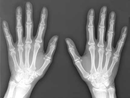
X-ray (Radiography) of Bone
A bone x-ray makes images of any bone in the body, including the hand, wrist, arm, elbow, shoulder, foot, ankle, leg (shin), knee, thigh, hip, pelvis or spine.
Common Uses
Preperation
Equipment
Procedure
Experience
Result Reading
Benefits vs. Risks
Limitations
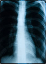
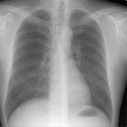
X-ray (Radiography) of Chest
The chest x-ray is the most commonly performed diagnostic x-ray examination. A chest x-ray makes images of the heart, lungs, airways, blood vessels and the bones of the spine and chest.
Common Uses
Preperation
Equipment
Procedure
Experience
Result Reading
Benefits vs. Risks
Limitations
Ultrasound
Ultrasound imaging, also called ultrasound scanning or sonography, involves the use of a small transducer and ultrasound gel to expose the body to high-frequency sound.
Common Uses
Preperation
Equipment
Procedure
Experience
Result Reading
Benefits vs. Risks
Limitations
X-ray (Radiography) of Bone
- Diagnose fractured bones or joint dislocation.
- Demonstrate proper alignment and stabilization of bony fragments following treatment of a fracture.
- Guide orthopedic surgery, such as spine repair/fusion, joint replacement and fracture reductions.
- Look for injury, infection, arthritis, abnormal bone growths and bony changes seen in metabolic conditions.
- Assist in the detection and diagnosis of bone cancer.
- Locate foreign objects in soft tissues around or in bones.

Most bone x-rays require no special preparation.
You may be asked to remove some or all of your clothes and to wear a gown during the exam. You may also be asked to remove jewelry, removable dental appliances, eye glasses and any metal objects or clothing that might interfere with the x-ray images. Women should always inform their physician and x-ray technologist if there is any possibility that they are pregnant. Many imaging tests are not performed during pregnancy so as not to expose the fetus to radiation. If an x-ray is necessary, precautions will be taken to minimize radiation exposure to the baby.
See the Safety page (www.RadiologyInfo.org/en/safety/) for more information about pregnancy and x-rays.
A portable x-ray machine is a compact apparatus that can be taken to the patient in a hospital bed or the emergency room. The x-ray tube is connected to a flexible arm that is extended over the patient while an x-ray film holder or image recording plate is placed beneath the patient.
X-rays are a form of radiation like light or radio waves. X-rays pass through most objects, including the body. Once it is carefully aimed at the part of the body being examined, an x-ray machine produces a small burst of radiation that passes through the body, recording an image on photographic film or a special detector.
Different parts of the body absorb the x-rays in varying degrees. Dense bone absorbs much of the radiation while soft tissue, such as muscle, fat and organs, allow more of the x-rays to pass through them. As a result, bones appear white on the x-ray, soft tissue shows up in shades of gray and air appears black.
Until recently, x-ray images were maintained as hard film copy (much like a photographic negative). Today, most images are digital files that are stored electronically. These stored images are easily accessible and are frequently compared to current x-ray images for diagnosis and disease management.

You must hold very still and may be asked to keep from breathing for a few seconds while the x-ray picture is taken to reduce the possibility of a blurred image. The technologist will walk behind a wall or into the next room to activate the x-ray machine. You may be repositioned for another view and the process is repeated. Two or three images (from different angles) will typically be taken around a joint (knee, elbow or wrist).
An x-ray may also be taken of the unaffected limb, or of a child's growth plate (where new bone is forming), for comparison purposes.
When the examination is complete, you will be asked to wait until the radiologist determines that all the necessary images have been obtained.
A bone x-ray examination is usually completed within five to 10 minutes.
A bone x-ray examination itself is a painless procedure.
You may experience discomfort from the cool temperature in the examination room. You may also find holding still in a particular position and lying on the hard examination table uncomfortable, especially if you are injured. The technologist will assist you in finding the most comfortable position possible that still ensures x-ray image quality.
A radiologist, a physician specifically trained to supervise and interpret radiology examinations, will analyze the images and send a signed report to your primary care or referring physician, who will discuss the results with you.
Follow-up examinations may be necessary, and your doctor will explain the reason why another exam is needed. Sometimes a follow-up exam is done because a suspicious or questionable finding needs clarification with additional views or a special imaging technique. A follow-up examination may be necessary so that any change in a known abnormality can be monitored over time.
Follow-up examinations are sometimes the best way to see if treatment is working or if an abnormality is stable over time.
Benefits
- Bone x-rays are the fastest and easiest way for a physician to view and assess fractured bone and joint abnormalities, such as arthritis and spine injuries.
- X-ray equipment is relatively inexpensive and widely available in emergency rooms, physician offices, ambulatory care centers, nursing homes and other locations, making it convenient for both patients and physicians.
- Because x-ray imaging is fast and easy, it is particularly useful in emergency diagnosis and treatment.
- No radiation remains in a patient's body after an x-ray examination.
- X-rays usually have no side effects in the typical diagnostic range for this exam.
- There is always a slight chance of cancer from excessive exposure to radiation. However, the benefit of an accurate diagnosis far outweighs the risk.
- The effective radiation dose for this procedure varies. See the Safety page (www.RadiologyInfo.org/en/safety/) for more information about radiation dose.
- Women should always inform their physician or x-ray technologist if there is any possibility that they are pregnant. See the Safety page (www.RadiologyInfo.org/en/safety/) for more information about pregnancy and x-rays.
While x-ray images are among the clearest, most detailed views of bone, they provide little information about muscles, tendons or joints.
An MRI may be more useful in identifying ligament tears and joint effusions in knee or shoulder injuries and in imaging the spine, because both the bones and the spinal cord can be evaluated. MRI can also detect a bone bruise when no crack is visible on x-ray images.
CT is being used widely to assess trauma patients in emergency departments. A CT scan can image complicated fractures, subtle fractures or dislocations. In elderly or patients with osteoporosis, a hip fracture may be clearly seen on a CT scan, while it may be barely seen, if at all, on a hip x-ray.
For suspected spine injury, 3-D reconstructed CT images can be made without additional radiation exposure to help the diagnosis and treatment of the individual patient's condition.
X-ray (Radiography) of Chest
A chest x-ray is typically the first imaging test used to help diagnose symptoms such as:
- Shortness of breath.
- A bad or persistent cough.
- Chest pain or injury.
- Fever. Physicians use the examination to help diagnose or monitor treatment for conditions such as:
- Pneumonia.
- Heart failure and other heart problems.
- Emphysema.
- Lung cancer.
- Line and tube placement.
- Other medical conditions.
A chest x-ray requires no special preparation.
You may be asked to remove some or all of your clothes and to wear a gown during the exam. You may also be asked to remove jewelry, removable dental appliances, eye glasses and any metal objects or clothing that might interfere with the x-ray images. Women should always inform their physician and x-ray technologist if there is any possibility that they are pregnant. Many imaging tests are not performed during pregnancy so as not to expose the fetus to radiation. If an x-ray is necessary, precautions will be taken to minimize radiation exposure to the baby.
See the Safety page (www.RadiologyInfo.org/en/safety/) for more information about pregnancy and x-rays.
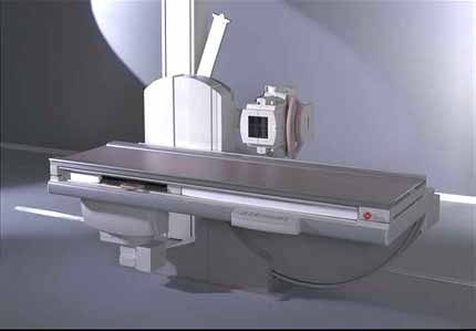
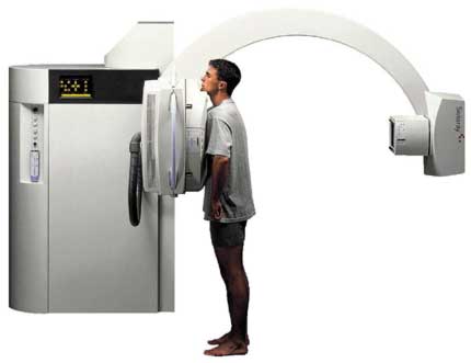
The equipment may also be arranged with the x-ray tube suspended over a table on which the patient lies. A drawer under the table holds the x-ray film or digital recording plate.
A portable x-ray machine is a compact apparatus that can be taken to the patient in a hospital bed or the emergency room. The x-ray tube is connected to a flexible arm that is extended over the patient while an x-ray film holder or image recording plate is placed beneath the patient.
X-rays are a form of radiation like light or radio waves. X-rays pass through most objects, including the body. Once it is carefully aimed at the part of the body being examined, an x-ray machine produces a small burst of radiation that passes through the body, recording an image on photographic film or a special detector.
Different parts of the body absorb the x-rays in varying degrees. Dense bone absorbs much of the radiation while soft tissue, such as muscle, fat and organs, allow more of the x-rays to pass through them. As a result, bones appear white on the x-ray, soft tissue shows up in shades of gray and air appears black.
On a chest x-ray, the ribs and spine will absorb much of the radiation and appear white or light gray on the image. Lung tissue absorbs little radiation and will appear dark on the image.
Until recently, x-ray images were maintained as hard film copy (much like a photographic negative). Today, most images are digital files that are stored electronically. These stored images are easily accessible and are frequently compared to current x-ray images for diagnosis and disease management.
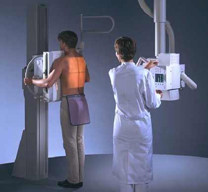
Typically, two views of the chest are taken, one from the back and the other from the side of the body as the patient stands against the image recording plate. The technologist, an individual specially trained to perform radiology examinations, will position the patient with hands on hips and chest pressed against the image plate. For the second view, the patient's side is against the image plate with arms elevated.
Patients who cannot stand may be positioned lying down on a table for chest x-rays.
You must hold very still and may be asked to keep from breathing for a few seconds while the x-ray picture is taken to reduce the possibility of a blurred image. The technologist will walk behind a wall or into the next room to activate the x-ray machine. When the examination is complete, you will be asked to wait until the radiologist determines that all the necessary images have been obtained.
The chest x-ray examination is usually completed within 10-15 minutes.
Additional views may be required within hours, days or months to evaluate any changes in the chest.
A chest x-ray examination itself is a painless procedure.
You may experience discomfort from the cool temperature in the examination room and the coldness of the recording plate. Individuals with arthritis or injuries to the chest wall, shoulders or arms may have discomfort trying to stay still during the examination. The technologist will assist you in finding the most comfortable position possible that still ensures diagnostic image quality.

The results of a chest x-ray can be available almost immediately for review by your physician.
Follow-up examinations may be necessary, and your doctor will explain the reason why another exam is needed. Sometimes a follow-up exam is done because a suspicious or questionable finding needs clarification with additional views or a special imaging technique. A follow-up examination may be necessary so that any change in a known abnormality can be monitored over time.
Follow-up examinations are sometimes the best way to see if treatment is working or if an abnormality is stable over time.
Benefits
- No radiation remains in a patient's body after an x-ray examination.
- X-rays usually have no side effects in the typical diagnostic range for this exam.
- X-ray equipment is relatively inexpensive and widely available in emergency rooms, physician offices, ambulatory care centers, nursing homes and other locations, making it convenient for both patients and physicians.
- Because x-ray imaging is fast and easy, it is particularly useful in emergency diagnosis and treatment.
- There is always a slight chance of cancer from excessive exposure to radiation. However, the benefit of an accurate diagnosis far outweighs the risk.
- The effective radiation dose for this procedure varies. See the Safety page (www.RadiologyInfo.org/en/safety/) for more information about radiation dose.
- Women should always inform their physician or x-ray technologist if there is any possibility that they are pregnant. See the Safety page (www.RadiologyInfo.org/en/safety/) for more information about pregnancy and x-rays.
The chest x-ray is a very useful examination, but it has limitations. Because some conditions of the chest cannot be detected on a conventional chest x-ray image, this examination cannot necessarily rule out all problems in the chest. For example, small cancers may not show up on a chest x-ray. A blood clot in the lungs, a condition called a pulmonary embolism, cannot be seen on chest x-rays.
Further imaging studies may be necessary to clarify the results of a chest x-ray or to look for abnormalities not visible on the chest x-ray.
Ultrasound
Ultrasound examinations can help to diagnose a variety of conditions and to assess organ damage following illness.
Ultrasound is used to help physicians evaluate symptoms such as:
- pain
- swelling
- infection Ultrasound is a useful way of examining many of the body's internal organs, including but not limited to the:
- heart and blood vessels, including the abdominal aorta and its major branches
- liver
- gallbladder
- spleen
- pancreas
- kidneys
- bladder
- uterus, ovaries, and unborn child (fetus) in pregnant patients
- eyes
- thyroid and parathyroid glands
- scrotum (testicles)
- brain in infants
- hips in infants
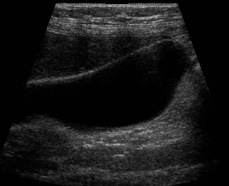
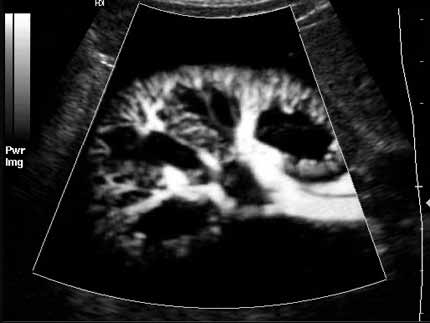
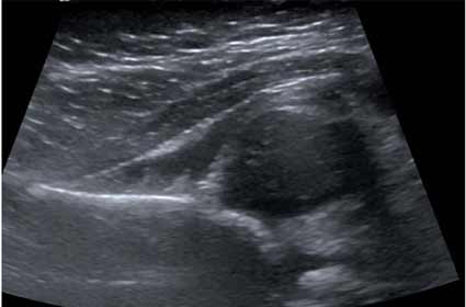
Ultrasound: Liver
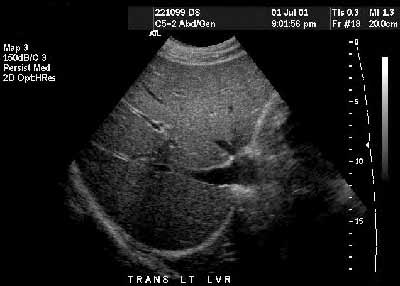
- guide procedures such as needle biopsies, in which needles are used to extract sample cells from an abnormal area for laboratory testing.
- image the breasts and to guide biopsy of breast cancer (see the Ultrasound-Guided Breast Biopsy page (www.RadiologyInfo.org/en/info.cfm?pg=breastbius)).
- diagnose a variety of heart conditions and to assess damage after a heart attack or diagnose for valvular heart disease. Doppler ultrasound images can help the physician to see and evaluate:
- blockages to blood flow (such as clots).
- narrowing of vessels.
- tumors and congenital vascular malformation.
With knowledge about the speed and volume of blood flow gained from a Doppler ultrasound image, the physician can often determine whether a patient is a good candidate for a procedure like angioplasty.
You should wear comfortable, loose-fitting clothing for your ultrasound exam. You may need to remove all clothing and jewelry in the area to be examined.
You may be asked to wear a gown during the procedure.
Other preparation procedures depend on the type of examination you will have. For some scans your doctor may instruct you not to eat or drink for as many as 12 hours before your appointment. For others you may be asked to drink up to six glasses of water two hours prior to your exam and avoid urinating so that your bladder is full when the scan begins.
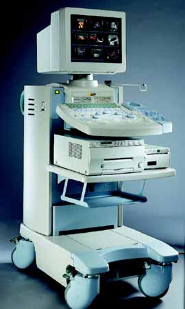
The ultrasound image is immediately visible on a video display screen that looks like a computer or television monitor. The image is created based on the amplitude (loudness), frequency (pitch) and time it takes for the ultrasound signal to return from the area of the patient being examined to the transducer, as well as the composition of body tissue through which and the type of body structure the sound travels through.
Ultrasound imaging is based on the same principles involved in the sonar used by bats, ships and fishermen. When a sound wave strikes an object, it bounces back, or echoes. By measuring these echo waves, it is possible to determine how far away the object is and its size, shape and consistency (whether the object is solid or filled with fluid).
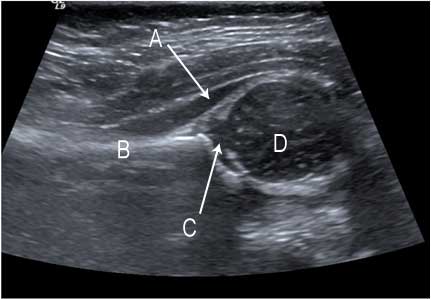
In medicine, ultrasound is used to detect changes in appearance of organs, tissues, and vessels or detect abnormal masses, such as tumors.
In an ultrasound examination, a transducer both sends the sound waves and receives the echoing waves. When the transducer is pressed against the skin, it directs small pulses of inaudible, high-frequency sound waves into the body. As the sound waves bounce off of internal organs, fluids and tissues, the sensitive microphone in the transducer records tiny changes in the sound's pitch and direction. These signature waves are instantly measured and displayed by a computer, which in turn creates a real-time picture on the monitor. One or more frames of the moving pictures are typically captured as still images. Small loops of the moving “real time” images may also be saved.
Doppler ultrasound, a special application of ultrasound, measures the direction and speed of blood cells as they move through vessels. The movement of blood cells causes a change in pitch of the reflected sound waves (called the Doppler effect). A computer collects and processes the sounds and creates graphs or color pictures that represent the flow of blood through the blood vessels.
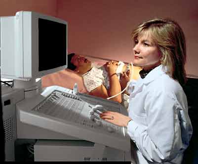
After you are positioned on the examination table, the radiologist or sonographer will apply a warm water-based gel to the area of the body being studied. The gel will help the transducer make secure contact with the body and eliminate air pockets between the transducer and the skin that can block the sound waves from passing into your body. The transducer is placed on the body and moved back and forth over the area of interest until the desired images are captured.
There is usually no discomfort from pressure as the transducer is pressed against the area being examined. However, if scanning is performed over an area of tenderness, you may feel pressure or minor pain from the transducer.
Doppler sonography is performed using the same transducer.
Children may need to be sedated in order to hold still for the procedure. Parents should ask about this beforehand and be made aware of food and drink restrictions that may be needed prior to sedation.
Once the imaging is complete, the clear ultrasound gel will be wiped off your skin.
In some ultrasound studies, the transducer is attached to a probe and inserted into a natural opening in the body. These exams include:
- Transesophageal echocardiogram. The transducer is inserted into the esophagus to obtain images of the heart.
- Transrectal ultrasound. The transducer is inserted into a man's rectum to view the prostate.
- Transvaginal ultrasound. The transducer is inserted into a woman's vagina to view the uterus and ovaries.
Ultrasound exams in which the transducer is inserted into an opening of the body may produce minimal discomfort.
If a Doppler ultrasound study is performed, you may actually hear pulse-like sounds that change in pitch as the blood flow is monitored and measured.
Most ultrasound examinations are completed within 30 minutes to an hour.
When the examination is complete, you may be asked to dress and wait while the ultrasound images are reviewed.
After an ultrasound examination, you should be able to resume your normal activities immediately.
Follow-up examinations may be necessary, and your doctor will explain the reason why another exam is needed. Sometimes a follow-up exam is done because a suspicious or questionable finding needs clarification with additional views or a special imaging technique. A follow-up examination may be necessary so that any change in a known abnormality can be monitored over time.
Follow-up examinations are sometimes the best way to see if treatment is working or if an abnormality is stable over time.
Benefits
- Most ultrasound scanning is noninvasive (no needles or injections).
- Occasionally, an ultrasound exam may be temporarily uncomfortable, but it is almost never painful.
- Ultrasound is widely available, easy-to-use and less expensive than other imaging methods.
- Ultrasound imaging is extremely safe and does not use any ionizing radiation.
- Ultrasound scanning gives a clear picture of soft tissues that do not show up well on x-ray images.
- Ultrasound is the preferred imaging modality for the diagnosis and monitoring of pregnant women and their unborn babies.
- Ultrasound provides real-time imaging, making it a good tool for guiding minimally invasive procedures such as needle biopsies and needle aspiration.
- For standard diagnostic ultrasound, there are no known harmful effects on humans.
Ultrasound waves are disrupted by air or gas; therefore ultrasound is not an ideal imaging technique for air-filled bowel or organs obscured by the bowel. In most cases, barium exams, CT scanning, and MRI are the methods of choice in this setting. Large patients are more difficult to image by ultrasound because greater amounts of tissue attenuates (weakens) the sound waves as they pass deeper into the body.
Ultrasound has difficulty penetrating bone and, therefore, can only see the outer surface of bony structures and not what lies within (except in infants who have more cartilage in their skeletons than older children or adults). For visualizing internal structure of bones or certain joints, other imaging modalities such as MRI are typically used.